Reperfusion Injury Risk Calculator
Risk Assessment Results
Quick Takeaways
- Reperfusion injury occurs when blood flow returns to tissue after a period of oxygen deprivation.
- Oxidative stress, calcium overload, and inflammation are the three core biological triggers.
- Targeted therapies - antioxidants, ischemic pre‑conditioning, and post‑conditioning - can cut damage by up to 40% in recent trials.
- Understanding the timing of interventions is crucial for heart attacks, strokes, and organ transplants.
- Staying up‑to‑date with 2024‑2025 research helps clinicians choose the right protocol for each patient.
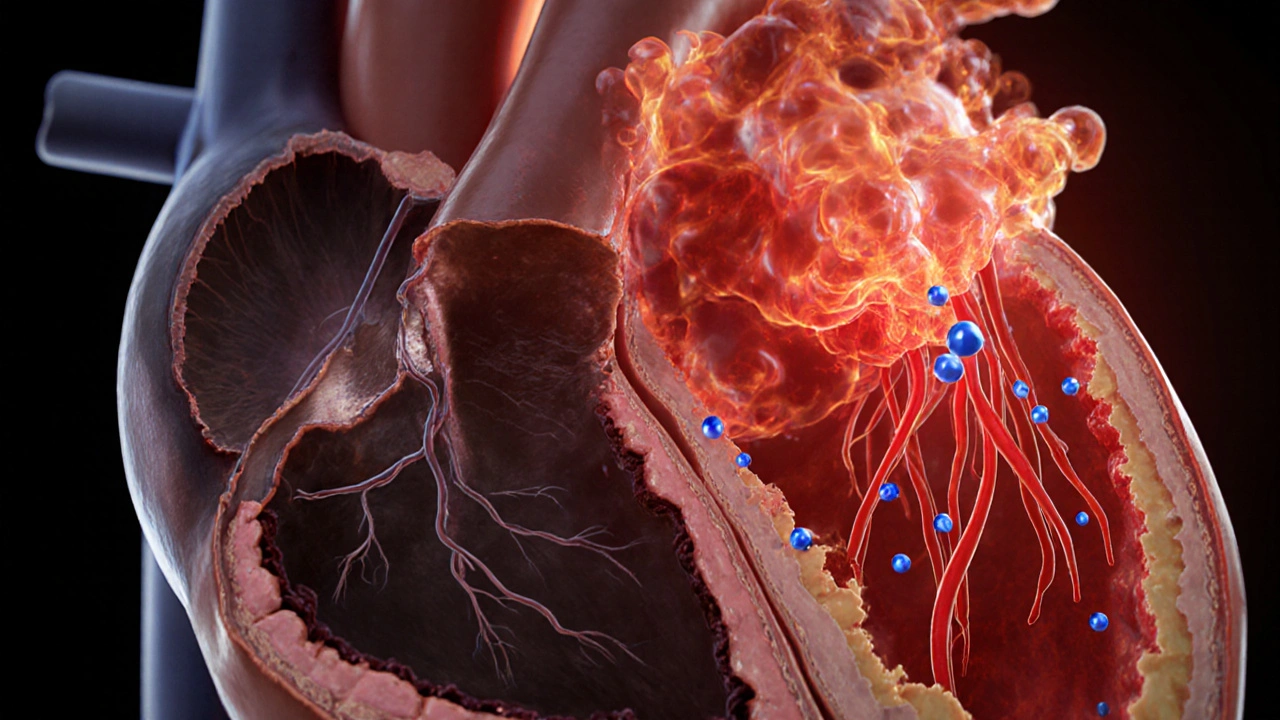
When blood rushes back into an area that’s been starved of oxygen, the sudden surge can backfire. The phenomenon, known as reperfusion injury is cellular damage that follows the restoration of blood flow after a period of ischemia, driven by oxidative stress, calcium dysregulation, and inflammatory cascades, and it underlies many of the complications seen after heart attacks, strokes, and organ transplants. This overview breaks down the science, highlights why the injury matters, and offers a practical checklist for reducing its impact.
What Is Reperfusion Injury?
In plain terms, reperfusion injury is the paradox of healing: the very act of “re‑oxygenating” a deprived organ creates a burst of harmful molecules that can destroy cells that were otherwise intact. The injury starts within seconds of blood returning and can continue for hours, shaping long‑term outcomes.
Why Does It Happen? The Core Mechanisms
Three interlocking pathways drive the damage:
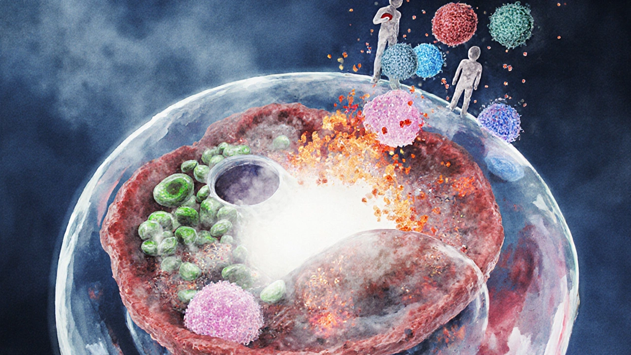
- Oxidative Stress - When oxygen floods back, mitochondria and enzymes generate massive amounts of reactive oxygen species (ROS). These free radicals oxidize lipids, proteins, and DNA, compromising cell membranes.
- Calcium Overload - The sudden shift in ion gradients forces calcium into cells. Excess calcium activates proteases and phospholipases that chew up structural proteins.
- Inflammatory Response - Re‑oxygenated tissue releases danger signals that attract neutrophils and macrophages. These immune cells release enzymes and more ROS, amplifying the injury.
All three feed into a final common pathway: the opening of the mitochondrial permeability transition pore (a channel that, when opened, collapses mitochondrial membrane potential, halting ATP production and triggering cell death). Once the pore opens, the cell can’t recover.
Key Players in the Damage Cascade
- Ischemia (a condition where blood flow-and therefore oxygen and nutrients-is insufficient to meet tissue demand)
- Reactive Oxygen Species (highly reactive molecules such as superoxide, hydrogen peroxide, and hydroxyl radicals formed during reperfusion)
- Calcium Overload (excess intracellular calcium that activates destructive enzymes)
- Inflammation (immune cell infiltration and cytokine release that worsen tissue damage)
- Mitochondrial Permeability Transition Pore (a protein complex that, when opened, leads to loss of mitochondrial integrity and cell death)
- Antioxidant Therapy (pharmacologic agents like N‑acetylcysteine or vitamin C that neutralize ROS)
- Ischemic Pre‑conditioning (brief, repeated cycles of ischemia that prime the heart to resist later injury)
- Clinical Outcomes (measurable patient results such as infarct size, ejection fraction, and neurological scores)
- Myocardial Infarction (the medical term for a heart attack, a primary setting where reperfusion injury is studied)
Clinical Impact: From Heart Attack to Stroke
Doctors first noticed reperfusion injury in patients undergoing thrombolysis or percutaneous coronary intervention (PCI) for myocardial infarction. Even when a blocked artery is reopened, up to 30% of the salvaged myocardium can be lost due to oxidative damage. In stroke care, the same paradox appears: rapid clot removal improves survival, yet the brain can suffer additional infarction from ROS and inflammation.
Organ transplantation offers another stark example. Donor organs endure cold ischemia, and when re‑warmer blood flows during implantation, reperfusion injury can trigger primary graft dysfunction, raising the risk of rejection.
Strategies to Reduce Reperfusion Injury
Researchers have tested a toolbox of interventions. Below is a snapshot of the most promising approaches, grouped by when they act.
| Timing | Intervention | Mechanism | Typical Efficacy (2023‑2025 trials) |
|---|---|---|---|
| Pre‑ischemic | Ischemic Pre‑conditioning | Triggers protective kinase pathways (e.g., PKC, Akt) | 30‑40% reduction in infarct size |
| Peri‑reperfusion | Antioxidant Therapy (N‑acetylcysteine, Vitamin C) | Scavenges ROS, restores glutathione | 15‑25% improvement in ejection fraction |
| Post‑ischemic | Ischemic Post‑conditioning | Interrupted blood flow bursts blunt ROS surge | 20‑35% smaller lesion volume in stroke models |
| Pharmacologic | Cyclosporine A (MPTP inhibitor) | Prevents mitochondrial pore opening | Modest 10‑12% functional improvement in MI patients |
| Cell‑based | Mesenchymal Stem Cell infusion | Modulates inflammation, releases trophic factors | Early trials show 22% better neurological scores |
Key takeaways:
- Timing matters more than the drug itself. A modest antioxidant given too late offers little benefit.
- Combining techniques (e.g., pre‑conditioning plus cyclosporine) yields additive protection in animal models.
- Patient‑specific factors-age, comorbidities, and genetic polymorphisms in antioxidant enzymes-affect response.
Latest Research Highlights (2023‑2025)
Several high‑impact studies have shifted the field:
- REDOX‑2024: A multicenter trial on intravenous edaravone (a free‑radical scavenger) showed a 28% reduction in left‑ventricular remodeling after primary PCI.
- PRE‑COND‑23: Demonstrated that a simple 5‑minute arm cuff inflation protocol before elective angiography cut cardiac enzyme release by 33%.
- MITO‑Guard 2025: Tested a novel peptide that selectively blocks the mitochondrial permeability transition pore; early phase‑II results indicated a 15% improvement in 90‑day functional scores after out‑of‑hospital cardiac arrest.
- Genomics work revealed that patients with the SOD2 Val16Ala variant experience 40% more oxidative damage, suggesting a future for personalized antioxidant dosing.
These findings reinforce the move toward tailored, timing‑precise protocols rather than one‑size‑fits‑all “antioxidant” pills.
Practical Checklist for Clinicians
- Identify high‑risk patients early (ST‑elevation MI, large‑vessel stroke, organ donor).
- Apply ischemic pre‑conditioning when feasible (e.g., brief cuff inflations) before reperfusion.
- Administer a fast‑acting antioxidant (edaravone or N‑acetylcysteine) within 5 minutes of restoring flow.
- Consider cyclosporine A or emerging MPTP inhibitors for eligible patients undergoing PCI.
- Monitor biomarkers: troponin, CK‑MB, and inflammatory markers (IL‑6, CRP) to gauge injury magnitude.
- Adjust therapy based on genetics if available (e.g., SOD2 genotype).
- Document outcomes (infarct size by MRI, ejection fraction, neurological scores) for quality improvement.
Following this flow helps turn the theoretical science of reperfusion injury into concrete bedside improvements.
Frequently Asked Questions
What exactly triggers oxidative stress during reperfusion?
When oxygen rushes back, enzymes in the mitochondria and NADPH oxidase rapidly produce reactive oxygen species. These free radicals attack cell membranes, DNA, and proteins, setting off the cascade that leads to cell death.
Can lifestyle changes reduce the risk of reperfusion injury?
Yes. Regular aerobic exercise improves endothelial function and boosts endogenous antioxidant defenses, which can blunt the ROS surge when blood flow is restored.
Is reperfusion injury only a concern for heart attacks?
No. It also plays a major role in strokes, peripheral artery disease interventions, and organ transplantation, whenever tissue experiences a period of ischemia followed by sudden re‑oxygenation.
How soon after reperfusion should antioxidants be given?
Evidence points to a 5‑minute window. Administering the drug within this brief period maximizes ROS scavenging before the bulk of the oxidative burst peaks.
Are there any risks associated with cyclosporine A for this purpose?
Cyclosporine can cause nephrotoxicity and hypertension, especially at higher doses. In the reperfusion setting, low‑dose regimens are used to balance pore inhibition with safety.
Understanding the science behind reperfusion injury equips clinicians, researchers, and even informed patients to make smarter choices when life‑saving blood flow is restored. By targeting oxidative stress, calcium overload, and inflammation at the right moment, we can turn a paradox into a pathway for recovery.
Earlene Kalman
Reading through the mechanisms, it’s clear that oxidative stress, calcium overload, and the inflammatory cascade are the three pillars of reperfusion injury. In practice, the 5‑minute window for antioxidant delivery is often missed because emergency teams are focused on restoring flow. The checklist at the end of the article could help keep that window top‑of‑mind.
Leon Wood
Hey everyone, great points! I love how the post breaks down the timing-getting those antioxidants in within minutes can really swing outcomes. For anyone on the front lines, think of it like a sprint: you’ve got to be fast, but also smart about which tools you use. Combining pre‑conditioning with a quick N‑acetylcysteine bolus is a win‑win.
George Embaid
Just to add a broader lens, remember that reperfusion injury isn’t just a U.S. issue; hospitals worldwide face the same timing hurdles. Incorporating culturally‑sensitive education for patients about the importance of early presentation can shave precious minutes off the door‑to‑balloon time. Sharing protocols across borders could lift overall success rates.
Meg Mackenzie
Honestly, I can’t shake the feeling that the mainstream medical community is being kept in the dark about the true scale of reperfusion damage. Every time they celebrate a new “miracle drug,” it’s really just a placebo wrapped in fancy statistics. The oxidative burst that follows reperfusion is like a hidden bomb, and the pharma giants profit from our ignorance. They push low‑dose antioxidants that barely cross the threshold needed to neutralize the ROS surge, then blame the failure on “patient variability.” Have you ever noticed how the trials that show big benefits are always sponsored by companies that own the patent? Meanwhile, independent labs that publish warning data get buried under paywalls and legal threats. The fact that the mitochondrial permeability transition pore is still not a primary target in most protocols is no accident; targeting it would mean admitting that we’ve been treating symptoms instead of the root cause. I’m also convinced that the “genetic tailoring” they talk about is a smokescreen to sell expensive genetic testing kits. The SOD2 variant mention feels like a marketing hook more than a genuine step toward personalized medicine. And let’s not forget the hidden costs of prolonged ICU stays caused by unchecked reperfusion injury-those dollars flow straight into the healthcare system’s bottom line. If we truly wanted to reduce mortality, we’d invest in rapid‑delivery antioxidant tech, not in endless conferences and blank‑paper publications. The article’s checklist is a good start, but without systemic change, it’s just a Band‑Aid on a bleeding wound. So, while the science looks solid on paper, the real‑world implementation is being sabotaged by conflicting interests. Stay vigilant, question the “standard of care,” and push for transparency. The truth is out there, buried under layers of corporate jargon.
Matt Miller
One practical tip: keep a pre‑filled syringe of N‑acetylcysteine in the cath lab so you can inject it the second the guidewire crosses the lesion. It saves seconds and cuts down on decision fatigue.
Barry White Jr
Timing is everything, no doubt.
Henry Kim
I appreciate the thorough breakdown, especially the part about monitoring biomarkers like IL‑6 and CRP. Those indicators can signal when the inflammatory wave is peaking, guiding us on when to add anti‑inflammatory agents. It’s also helpful to note that the checklist can be turned into a simple bedside app for quick reference.
Neha Bharti
From a coaching perspective, encouraging patients to adopt regular aerobic exercise pre‑emptively builds up their endogenous antioxidant systems, making the reperfusion shock less severe. Think of it like training for a marathon; the body becomes more resilient to stressors.
Samantha Patrick
Hey folks, just a quick heads‑up – the article forgets to mention that you should double‑check the IV line for any air bubbles before the antioxidant push. Those tiny bubbles can cause micro‑emboli, which adds another layer of injury.
Ryan Wilson
While I respect the enthusiasm, let’s not forget that pushing every patient through aggressive antioxidant protocols without clear evidence can be risky. Some studies have shown renal side effects with high‑dose N‑acetylcysteine, especially in people with underlying kidney issues. It’s essential to weigh benefits against potential harms and not treat every case with a one‑size‑fits‑all mindset.